
 Life Sciences
 Information Communication
 Environment
 Nanotechnology / Materials
 Energy
 Manufacturing Technology
 Social Infrastructure
 Frontier
 Human and social sciences

 Visualization of Biological Microstructure with High Frequency Ultrasound and Photoacoustic Imaging
update:2020/06/16
- Features and Uniqueness
-
- "Features"
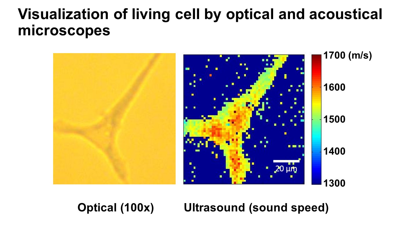
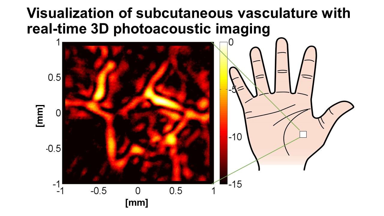
- High-resolution imaging of biological tissue is non-invasively obtained with high frequency ultrasound. We have developed some ultrasound microscope systems which realized the resolution of 15-micron with 100 MHz and resolution to visualize a single cell with GHz range ultrasound. Ultrasonic imaging provides not only tissue morphology but also information on tissue elasticity. Recently, we have developed a real-time three-dimensional photoacoustic imaging system for visualization of subcutaneous micro vasculature and oxygen saturation.
- "Targeted Application(s)/Industry"
- High frequency ultrasound and photoacoustic imaging is repeatedly and non-invasively applied for early diagnosis of atherosclerosis, skin aging and tissue metabolism. They are useful for efficacy assessment of cosmetics and pharmaceuticals. High frequency ultrasound is also applied in the industrial areas where thickness measurement of opaque film or bilayer thin coating with the precision of 0.1 micron is required.
- Keywords
Researchers
Graduate School of Biomedical Engineering
Yoshifumi Saijo, Professor
PhD (Medicine)
